Ultrasonic studies for a better society and humanity
Ultrasound is defined as “sound waves at frequencies beyond the auditory range of humans.” In recent years, however, ultrasound has been used in research unrelated to hearing, as “ultrasound technology” without reference to frequency. Ultrasound penetrates invisible objects, enabling “nondestructive” examination of their interior. Its propagation behavior changes with various physical properties and quantities, including magnetic field, temperature, elasticity, etc. “Ultrasound electronics” is the term used for all types of basic and applied research which involve ultrasound techniques. This research field is considered to be a fusion of various disciplines, including electronics and instrumentation engineering, mechanical engineering, chemistry, informatics, medicine, biology, etc.
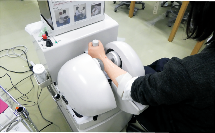
Dr. Matsukawa’s aim is to create technologies that will be useful for society. In the field of ultrasound electronics, she focuses on research and development, especially for opto-acoustics technology and medical applications. This includes the rather rare field of medical ultrasound “bone evaluations”. While, currently, ultrasonic diagnostic equipment is chiefly used in medical diagnosis, as often experienced in general medical checkups, the target in this field is “soft tissue.” A little progress has been made on ultrasonic diagnosis of bones because acoustic waves almost reflect from the surfaces of hard objects like bones and thus do not enter well. For the diagnosis of bones, X-ray techniques are the gold standard. However, these techniques suffer from problems due to radiation exposure, which is not good for children and pregnant women. In recent years, bone evaluations have been made for various purposes such as bone growth in children. Dr. Matsukawa has performed a large-scale study of junior and senior-high school students of Kyoto Prefecture in collaboration with Kyoto Prefectural University of Medicine. For this study, Dr. Matsukawa used an ultrasonic bone-density measurement apparatus which can evaluate, at accuracy rates similar to those obtained with computed tomography (CT), each portion of the radius of the wrist. (This device was manufactured by Oyo Electric Col, Ltd., and was jointly developed by Horiba, Ltd., and Doshisha University (Photo 1).)

Data obtained from more than 1300 individuals have been used to report novel results. This can also contribute to health guidance and advice for youth in their teens, including differences in bone growth due to sex and lifestyles. The effects of different dietary practices are also discussed.
Another problem indicated in recent years is bone fracture due to diabetes. Dr. Matsukawa has successfully reported the effects of diabetes on the ultrasonic wave properties in bone. Her unique approach using an opto-acoustic technique has enabled the measurements of ultrasonic wave velocity in the minute area of a bone. Contrary to the expectation, her group has found small velocity decrease in the diabetic bone, where non-physiological crosslinks such as advanced glycation end products exist. This means that diabetic bones are a little softer than normal bones.
Dr. Matsukawa states: “Ultrasonic studies have deepened our understanding of bone properties. Recently, we have been focusing not only on diagnosis but also on ultrasonic treatment.” Already, ultrasonic bone-fracture healing has become a common medical care. Several clinical reports have reported how ultrasonic irradiation can reduce the treatment period for fractures, and how it can be used in the treatment of refractory (i.e., difficult to cure) bone fractures. It seems, however, that the mechanisms of ultrasonic bone-fracture healing are yet to be clarified perfectly. For example, why does ultrasound irradiation enable rapid bone healing, and what are the effects of high-frequency pressure on cells? Dr. Matsukawa continues her pursuit to elucidate these mechanisms, with a special focus on electromagnetic phenomena which occur in bones under ultrasound irradiation.
Challenging original research works
Dr. Matsukawa pursues themes of interest where other researchers are not well engaged, and this means that she regularly crosses research boundaries. As for bone studies, she is also developing ultrasonic techniques that will enable early detection of leg-bone diseases in racehorses. “I started this research when a researcher from the Equine Research Institute of the Japan Racing Association (JRA) approached me at a domestic conference. Training of racehorses begins when they are around two years old. However, periostitis, a bone inflammation, can occur in the bone of a horse’s leg, and is known as ‘bucked shin.’ Sometimes, it can be so painful that the affected horse can no longer run. Performing X-ray examination is difficult for a large animal like a horse. Then I wondered if we could modify the ultrasonic diagnostic apparatus used for human bones for the early screening of bucked shin even outdoors in the field.” Her first paper on this issue was published in the Journal of the Acoustical Society of America and garnered great interest. “It was known that a modified human ultrasonic diagnostic machine could be used to evaluate horse muscles, however, the use of ultrasound to analyze the most important bones for running was unknown. It was very interesting.”
Dr. Matsukawa is also developing different research projects. She is diligently involved in the development of new sensor systems for detecting ultrasonic waves using surface plasmon resonance. In recent years, she has also performed joint research with private companies, medical institutions, etc., on methods of evaluating cerebral blood vessels. It is currently possible to use a medical ultrasonic diagnostic apparatus to evaluate vessels like carotid arteries, major blood vessels that supply blood and oxygen to the brain. Nevertheless, it is difficult for a family doctor to evaluate blood vessels within the brain that are surrounded by the hard skull bone.

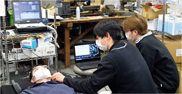
Moreover, only people with proper qualifications (like medical doctors and clinical technicians) are permitted to use diagnostic machinery like CT and MRA in the hospital. Thus, Dr. Matsukawa came up with the idea for simple measurements of carotid arterial pulse waves using a low-cost ultrasonic apparatus (Photo 3).

This enables the measurement of characteristic changes in the pulse waves of patients with cerebral artery occlusion and other cerebrovascular disorders, like placing the palm of the hand on the carotid artery. “Only a limited number of hospitals are able to treat serious issues like cerebral infarction, occlusion, and aneurysm. Without early treatment, it becomes difficult to recover from these diseases and may even cost live. In the field of emergency medicine, it is difficult to determine the specific ailment. Our goal is to develop a cerebral artery screening device that is safe and cheap. If we can screen the patient in the emergency medicine, the patient is sent to the appropriate hospital as quickly as possible. We are also considering the use of AI and other technologies to assist in the diagnosis of occlusion using pulse wave information obtained via measurement with ultrasonic techniques. We have recently started collaborations with researchers (professors, etc.) in information technology-related fields.”
Participation in joint research of all types: domestic and international, multidisciplinary, with industry, and with academia
Dr. Matsukawa has been pursuing joint research with overseas collaborators, centered in France, the country where ultrasound research began. She says that her motivation is the new perspectives she gains as she collaborates with researchers and engineers from a variety of fields. “Conversations with people in information-related fields have changed the ways I look at data, while my interactions with medical specialists have brought unexpected surprises and enlightenment. Joint research is truly stimulating.”
Dr. Matsukawa emphasizes the importance of collaborations not only within academia, but also with industry. She says that she has enjoyed sustained relationships over many years with companies in the ultrasound field. “Discussion and debate that includes researchers, students, company engineers, and all kinds of individuals is exciting, fascinating, and motivating. I often imagine how wonderful it would be not only to have the ‘high-level’ ultrasound equipment used in hospitals and medical exams but also if we had ultrasound screening systems that could be used by doctors and other medical specialists—and even in our own homes! ‘Sound’ is a physical phenomenon familiar to everyone and is taught in the elementary school. I hope that everyone can gain a better understanding of how important and helpful ‘sound’ is to all of us.” Ultrasound is currently in active use in numerous fields, not only in medical care but also for sonar, filters, nondestructive evaluations, motors, and more. Dr. Matsukawa would like to see the amazing developments yet to come in ultrasound studies.
