
Interview with researchers 17
Aiming for the elucidation
of brain functions
and medical applications
via creative technological development
—Observation of active brain functions
and the development of neuronal fibers
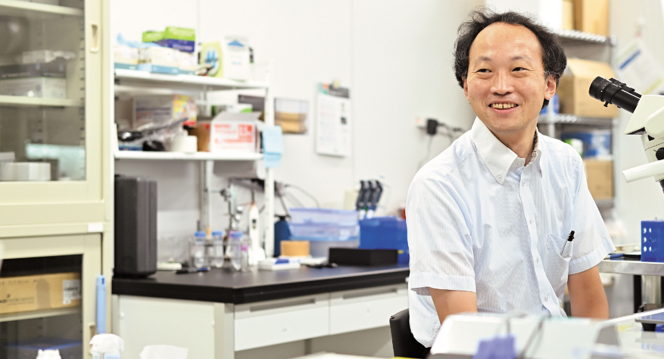
- Interview Yoshito Masamizu
- Professor,
Graduate School of Brain Science
Observing brain activities with the aim of illuminating its functions
Neurophysiology is the scientific domain that investigates brain activities, including memory and learning. The aim is to determine brain-cell connection circuits (neurocircuits). Since “brain functions” is a broad term, this domain encompasses a wide range of fields, from basic psychology to cognitive neuroscience.
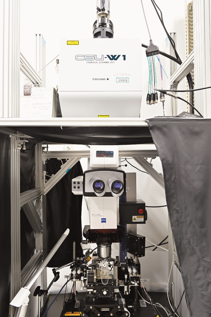
The development of observation methods is indispensable for the growth of this research domain. The key is to be able to capture the “real-time” state of the brain in more and more detail while it is actively working. Making those parts of the brain that are still largely unexplained visible is linked with the finding of novel facts. Dr. Masamizu, who specializes in neurophysiology, uses in vivo calcium imaging, a technique established around 2005, to explore and discover the true nature of the brain and its numerous mysteries. “We look at areas of the brain performing processing when an animal is performing a cognitive task, to determine how its brain cells (neurons) are acting. In in vivo calcium imaging, fluorescent calcium sensor proteins are used to monitor neural activity, which we then observe using a two-photon microscope and a macro zoom/multipoint laser scanning confocal microscope (Photo 1). This in vivo calcium imaging makes it easy to observe long-term activity changes in the same neurons. It is thus excellent for observing brain activity in animals used to perform experiments. Using this technique, we are clarifying neurocircuit networks involved in learning and memory, and more.”
Dr. Masamizu has been fascinated with the brain since his childhood. He selected a career as an academic researcher in order to learn and help unlock the principles of the brain. In his years as a graduate student, his research focused on the specialized field of neuroembryology, which studies the formation and development of the brain from the earliest stages. After he received his Ph.D., he moved to the field of neurophysiology. “I had a strong desire to know more and more about the brain, and thus studied brain development processes. However, as I pursued my research, I became aware of the need to know how the brain acts in order to acquire brain functions. This was the spark that led to my moving on to neurophysiology.”
Edging closer to new brain facts through step-by-step technological development
Dr. Masamizu uses in vivo calcium imaging to study rodents and primates. Their team was the first worldwide to determine that memory of learned movements is stored in a deep layer of the cerebral cortex. “The cortex has a six-layer structure. Prior research had observed layer 2/3 nearest the surface using in vivo calcium imaging. We made the special arrangement, which enabled the specific expression of fluorescent calcium sensors, used in in vivo calcium imaging, in deep layers. This enabled us to make observations of activity in deeper layers than before. As a movement task for a mouse, we devised an experiment where the mouse would receive water when pulling a lever for a fixed time. As result, we discovered that, in the late stage of learning, there was unique activity within layer 5 of the cerebral cortex motor area. We suggested that the neurons there might be responsible for motor-skills memory.”
Dr. Masamizu aims to reveal more about human brain functions. Using the marmoset—a primate that more closely resembles humans—to perform movement task experiments, he has also monitored neuronal activities in the marmoset’s cerebral cortex. “The marmoset is a comparatively small primate, and it was the first time for us to perform head-fixed movement-task experiments using this animal. The marmoset is especially sensitive to stress, so there is always a fear that it will stop performing movement task. We repeatedly had to go through numerous technical trial-and-errors before we successfully performed an experiment.” What contributed greatly to this success was the training apparatus specifically designed and developed for marmoset use, and a training method which involved making the training more difficult precisely in tandem with the extent that the marmoset became used to the assigned task. Especially useful was the idea to have the marmoset wear a specially designed training “jacket,” which enabled smoother training performance. Dr. Masamizu discovered this while reading a scientific paper outside his field, related to medical-drug administration. The jacket was realized by Dr. Masamizu’s technical assistants on the basis of illustrations provided in the text. “Working with an animal that had no manual entailed step-by-step changes and improvements along the way. We are confident that our results are the first step in a new direction for elucidating the mechanisms of human cognition and movement, as well as the fundamentals of neuronal networks.”
Neuronal fibers, rich in potential for novel developments in a range of fields, from medical application to artificial intelligence
While Dr. Masamizu has continued with his basic research to pinpoint healthy brain activities, he is also challenging applied research. This includes the creation of neuronal fibers, plus the establishment of technologies for the implantation of these fibers into the brain for functional recovery. An ordinary neuron has one extension called the axon (as well as others called dendrites). The axon extends in a specific direction, and exchanges information with other neurons. When existing neurons are damaged in their original site, they lose their functions, as the axons do not extend in their specific directions. This has made the transplants of neuronal cell suspensions very difficult. Dr. Masamizu seeks to create neuronal fibers that maintain their orientation and can be transplanted within the brain. Currently, brain recovery is difficult when neurons are transplanted into areas in the brain damaged due to stroke, etc., especially depending on the width of the transplant area and how much time has elapsed since the damage occurred. In the future, if Dr. Masamizu can successfully develop neuronal fibers, it is hoped that a transplant that enables a bypass (detour) of the damaged area could lead to brain recovery (Fig. 1).

Fig. 1 Image diagram of bypassing damaged areas using the transplant of the neuronal fiber
If the implant of the neuronal fiber becomes possible, damaged brain areas beyond recovery can be bypassed, and new neurocircuits can be created. This could enable recovery of brain functions.
[Left image] In the case where information flows in the order A → B → C, when B is damaged, C cannot function.
[Right image] With the transplant of the neuronal fiber, B can be avoided, and a novel neuronal circuit is created
Dr. Masamizu also seeks ways to guide new discoveries with his research using in vivo calcium imaging. “We are working on a new approach, and that is to create new circuits in the brain with the transplantation of neuronal fibers, as well as to observe brain activity during that approach using the in vivo calcium imaging techniques that we have previously developed and refined. Recent developments in artificial technology research have also been performed using hints and suggestions from actual brain activity. With the construction of novel neuronal circuits, if we can see changes in brain functions as a result, it should be easy to incorporate these within mathematical models. We look forward to making contributions to this research area as well.”
Dr. Masamizu and his lab have thus continued to generate new and productive results in both basic and applied research. He says that his motivations for research have spurred his continued movement along paths that are not always the accepted and “correct” ways. “The brain is extremely complicated, and searching within its dark passageways can often be difficult and frustrating. But it is its unpredictability that makes experimentation and measurement, as well as making novel discoveries, so interesting and worthwhile.” Dr. Masamizu continues to perform his challenging work to unlock and solve puzzles of the brain. We can all eagerly look forward to more new and exciting research discoveries from him and his team into the future.
PROFILE
- Research Area
- Neurophysiology
- Research Themes
- • Establishment of a system for brain transplants of neuronal fibers
• Neuronal circuit construction for recovery and extension of brain functions
- Research Goals
- (1) Establishment of a system for brain transplants of neuronal fibers
(2) Neuronal circuit construction for recovery and extension of brain functions
- Researcher Database
- https://kendb.doshisha.ac.jp/profile/en.b1bfc169e35e4722.html