
Research News: Imaging Epigenetic Mechanisms that Help Bone Resorbing Cells Handle Oxygen Stress
November 24, 2021
Scientists in Japan develop novel microscopy to analyze oxygenation within living murine bone tissue, with implications for bone resorption
With futuristic life science research comes the challenge of monitoring biological parameters in live animals. Taking the leap forward are scientists from Doshisha University who have developed an advanced imaging technique to measure oxygen concentration in live animal tissues. They were able to measure oxygenation in osteoclasts—giant cells involved in bone resorption—under different conditions, and identify the route of osteoclastogenesis that involves oxygen-mediated DNA methylation within the physiological range of oxygen tension.
Without doubt, the future of life sciences is sure to have technology that can measure biological parameters real-time in living organisms. This would definitely help monitor tissue homeostasis, on the go. One important component of such homeostasis is the tissue oxygen concentration.
Under normoxic conditions, the tissues have enough oxygen for the upkeep of biological functions. But in hypoxic conditions—the state of oxygen deprivation inducible in real life during travels at high altitudes or through asphyxiation—the tissue may not have enough oxygen for such upkeep. This triggers a hypoxic response. The cells involved are likely to activate mechanisms that ensure hypoxia mitigation.
To explore further the intricacies of cellular response to variable oxygen concentrations in the living body, as a first futuristic step, scientists from Doshisha University, Kyotanabe, have successfully developed an advanced imaging technique involving two-photon phosphorescence lifetime imaging microscopy. They applied this microscopy technique in live mice to measure oxygen concentration in situ within multinucleated giant cells called osteoclasts that facilitate bone resorption. Their findings have been published as a research article in EMBO Reports.
Professor Keizo Nishikawa of Graduate School of Life and Medical Sciences, Doshisha University, the lead author of the study, explains the motivation behind their research, thus: “Through this study, we suggest a new approach to understand the physiological mechanisms of living organisms by accurately measuring the concentration of biomolecules deep in biological tissues. We believe ours is the first quantitative research attempt in the life sciences, similar to the that applied in the physical and chemical sciences to understand natural phenomena mathematically.”
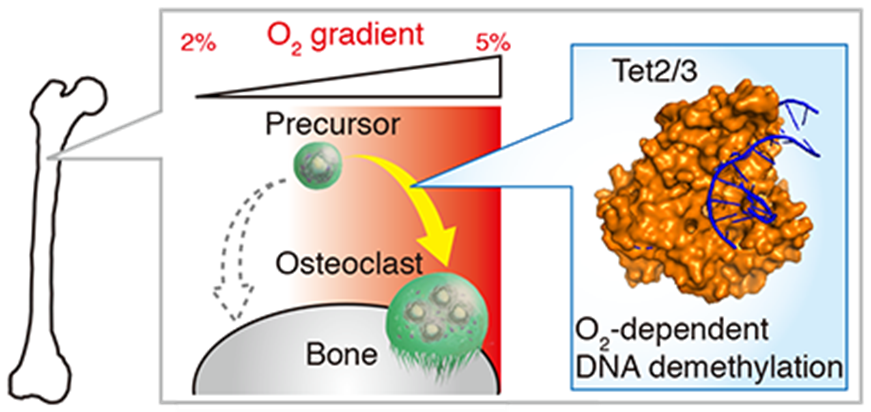
Accordingly, through their work, the scientists were able to determine the physiological range of oxygen tension within osteoclasts of live mice, which they note to be from 17.4 to 36.4 mmHg, under hypoxic and normoxic conditions, respectively. This means that physiological normoxia corresponds to 5% and hypoxia to 2% oxygen in osteoclasts.
Also, their independent analysis of hypoxic conditions in bone tissue showed that the dearth of oxygen severely limits osteoclastogenesis or the process by which new osteoclasts grow. Further, they were able to identify that hypoxia interferes with the activity of ten-eleven translocation (TET) enzymes, which play a vital role in osteoclastogenesis through oxygen-dependent DNA methylation. Thus, the scientists suggest that osteoclastogenesis may be limited independent of energy metabolism and hypoxia-inducible factor activity under conditions of 2% oxygen in osteoclasts.
Overall, the study has not only put forth a novel imaging technique to measure oxygen concentration but has also deciphered a key molecular mechanism involved in osteoclastogenesis. The importance of TET enzymes in regulating osteoclastogenesis within the physiological range of oxygen tension may help unravel hitherto unreported mechanisms of physiological response to oxygen perturbation. Professor Nishikawa further hopes, “We expect our findings to be applied in new medical technologies that predict disturbances of homeostasis and disease onset in the body.”
The importance of sensing of oxygen by cells and their adaptation to its availability received its due recognition through award of the 2019 Nobel Prize in Physiology or Medicine. The current research by scientists from Doshisha University promises to add to this increasing body of knowledge on what constitutes the quintessential cellular mechanism keeping us all alive.
Scientists from Japan develop advanced imaging technique to measure oxygenation in live mouse bone
Scientists from Doshisha University have used two-photon phosphorescence lifetime imaging microscopy to measure oxygen concentration within osteoclasts of live mice. Through live oxygen measurements, they have further identified a novel response through oxygen-mediated DNA methylation to elicit osteoclastogenesis.
Image courtesy: Keizo Nishikawa from Doshisha University
Reference
| Title of original paper | Osteoclasts adapt to physioxia perturbation through DNA demethylation |
| Journal | EMBO Reports |
| DOI | 10.15252/embr.202153035 |
| Latest Article Publication Date | 18th October 2021 |
| Method of Research | Experimental study |
| Subject(s) of Research | Animals |
| Conflict of Interest statement | The authors declare that they have no conflicts of interests |
Funding information
This study was financially supported by Grants-in-Aid for Scientific Research (B) from the Japan Society for the Promotion of Science (JSPS) (18H02614); Grants-in-Aid for Scientific Research on Innovative Areas from the JSPS (17H05530); CREST, Japan Science and Technology Agency; Grants-in-Aid for Scientific Research (S) from the JSPS (19H05657); Grants-in-Aid for Scientific Research on Innovative Areas from the JSPS (26111001 and 26111004); and grants from the Takeda Science Foundation, Toray Science Foundation, the Nakatani Foundation, Yamada Science Foundation, LOTTE Foundation, Suzuken Memorial Foundation; and Terumo Life Science Foundation. Also, the infrastructure of metabolomics was partly supported by JST ERATO Suematsu Gas Biology Project.
Profile
Dr. Keizo Nishikawa is a Professor at Faculty of Life and Medical Sciences Department of Medical Life Systems, in Doshisha University, Japan. Dr. Nishikawa’s research expertise spans broad areas of cell and molecular biology, pharmacology, medicinal biochemistry, food sciences, and laboratory animal sciences. In these fields, he has authored around 30 research papers, which has been published in national and international journals.
Keizo Nishikawa
Professor, Faculty of Life and Medical Sciences, Department of Medical Life Systems
Media contact
Organization for Research Initiatives & Development
Doshisha University
Kyotanabe, Kyoto 610-0394, JAPAN
CONTACT US