
Research News: Listening in on Neuron Conversations: Novel Fluorescent cAMP Indicator Facilitates Neuronal Live-Cell Imaging
July 14, 2022
Tracking intercellular dynamics in neurons using the novel fluorescent probe, gCarvi, may help decode neuronal talking.
Cyclic adenosine monophosphate (cAMP) is an essential signaling molecule. However, probes that provide a clear view of intracellular cAMP dynamics are yet to be developed. Now, scientists from Doshisha University have created a novel cAMP-specific fluorescent indicator called gCarvi, that displays cAMP concentration in neurons. They found that using gCarvi revealed information regarding cAMP’s multifaceted role in molecular signaling pathways. gCarvi could therefore be a powerful tool to determine cAMP dynamics underlying several neuronal functions.
Cyclic adenosine monophosphate (cAMP) is an intracellular messenger molecule responsible for the functioning of numerous cells, including neurons, where it promotes the growth of axons and maintains neuronal communication. The molecular pathways of cAMP are well studied and it is known to play a major role in regulating synaptic functions; however, indicators that can precisely monitor its intracellular activity are yet to be developed.
Now, in a study published in Proceedings of the National Academy of Sciences, a team of researchers led by Associate Professor Naoto Saitoh from Doshisha University solved this problem through their development of a novel green fluorescent cAMP indicator called gCarvi. Their indicator can be easily expressed using an adeno-associated virus vector and is capable of quantitatively measuring the basal concentration of cAMP (called [cAMP]i) intracellularly in neurons. “Our new indicator, gCarvi, monitors [cAMP]i at concentrations ranging from 0.2 to 20 μM with a subsecond time resolution and a high specificity over cGMP. It is bright enough to monitor cAMP kinetics in single cells and even at single presynaptic boutons,” explains Dr. Saitoh, who is the corresponding author of the study. His team included Ms. Seiko Kawata, a PhD student from Doshisha University and the first author of the study, and Professor Tomoyuki Takahashi from Okinawa Institute of Science and Technology. This paper was made available online on July 6, 2022 and was published in Volume 119 Issue 28 of the journal on July 12, 2022.
An ideal cAMP indicator must span the entire intracellular range of cAMP concentration, exhibit high specificity for cAMP over the other nucleotides such as cyclic guanosine monophosphate (cGMP) which is also responsible for synaptic modulation, not interfere with physiological functions of endogenous cAMP pathways, and enable stable intracellular imaging. Most existing cAMP indicators consist of regulatory subunits of the cAMP binding domain (CBD) in mammals. However, these subunits are endogenous molecules and their use could disrupt the physiological functions of cAMP pathways. gCarvi overcomes this problem as it contains the CBD of a bacterial cAMP receptor protein which does not interfere with the cAMP pathways in mammalian cells.
The researchers fused the bacterial CBD to a circular permutated green fluorescent protein (cpGFP), which detects cAMP concentration by causing corresponding changes in the intensity of fluorescent light emitted upon binding to it. Additionally, gCarvi was found to counter another setback of existing cAMP indicators in preventing cGMP detection by exhibiting a high cAMP/cGMP specificity of >100 but a low affinity to cGMP.
The team developed a ratiometric probe by fusing gCarvi with a red fluorescent protein called mCherry to improve its ability to detect various concentrations of cAMP. This probe was expressed in hippocampal neurons to monitor variations in [cAMP]ias a proof of concept.Similarly, gCarvi was co-expressed with a red fluorescent calcium ion (Ca2+) indicator to detect cAMP and Ca2+, both which play a role in neuronal regulation.
This simultaneous detection of cAMP/Ca2+ in the hippocampal neurons revealed a strong link between cAMP and Ca2+ signals. Upon treatment with forskolin, which is a compound that raises cAMP levels, and a phosphodiesterase (PDE) inhibitor, an increase in intracellular cAMP was observed above a certain threshold in these neurons, which resulted in Ca2+ elevation. Similarly, in cerebellar presynaptic boutons, forskolin was used to induce irregular cAMP elevations that demonstrated positive increases in the number of synaptic vesicles being recycled. This indicates the presence of presynaptic cAMP domains in individual boutons, which improve synaptic strength by compartmentalizing signaling pathways to avoid cross-talk.
Speaking of future applications of this novel indicator, Dr. Saitoh says “gCarvi can precisely detect the time of degradation of cAMP signaling in the brain, which results in neuro-degenerative conditions like Alzheimer’s disease. gCarvi can be employed as a screening tool in live-neuron imaging to monitor outcomes of drugs targeting such conditions. Additionally, fluorescent probes help detect invisible molecular dynamics in cells; therefore, the gCarvi cAMP indicator presents invaluable knowledge on neuronal talking.”
The future is certainly looking bright for gCarvi!
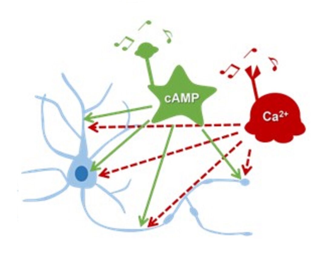
Cross-regulation between cAMP/Ca2+ is a fundamental phenomenon in neurons
Cross-regulation between cAMP/Ca2+ is a fundamental phenomenon in neurons. However, we do not know the precise details of cAMP dynamics. Now, researchers from Japan have developed a new fluorescent cAMP indicator that has led to invaluable insights on neuronal communication.
Image courtesy: Naoto Saitoh from Doshisha University
Image license: CC BY-NC-ND 4.0
Image link: https://wordpress.org/openverse/image/5e6026b8-a594-429f-981f-422747131c30
Reference
| Title of original paper | Green fluorescent cAMP indicator of high speed and specificity suitable for neuronal live-cell imaging |
| Journal | Proceedings of the National Academy of Sciences (PNAS) |
| DOI | https://doi.org/10.1073/pnas.2122618119 |
Funding information
This study was supported by the Core Research for Evolutional Science and Technology of the Japan Science and Technology Agency, Grant-in-Aid for Scientific Research from MEXT Japan, and the Doshisha Harris Grant.
Profile

Dr. Naoto Saitoh is an Associate Professor from the Faculty of Biomedical Sciences at Doshisha University. Dr. Saitoh holds a PhD in Agriculture from the University of Tokyo. He has held many notable research positions prior to joining Doshisha University, from being a CREST researcher at the Department of Functional Biology to being a specially appointed lecturer at the Department of Neurophysiology at the University of Tokyo. Dr. Saitoh has over 20 research publications to his credit with a research focus on Cellular and Molecular Neuroscience.
SAITOH Naoto
Associate Professor , Faculty of Life and Medical Sciences Department of Medical Life Systems
Media contact
Organization for Research Initiatives & Development
Doshisha University
Kyotanabe, Kyoto 610-0394, JAPAN
CONTACT US