Research News:Ultrasonication as a Tool for Directing Cell Growth and Orientation
December 05, 2024
Scientists develop ultrasound-based technique to control myoblast (cultured muscle cell) orientation, advancing tissue engineering and regenerative medicine
Developing reliable methods to replace damaged tissue remains a central goal in tissue engineering and regenerative medicine.Now, researchers in Japan have created an ultrasound-based technique to control the orientation of cultured myoblasts—precursor cells to skeletal muscle. This innovative method could enable the production of aligned cell sheets s/news-detail-62uitable for directtransplantation, offering new possibilities for therapeutic applications in regenerative medicine and muscle tissue engineering.
Developing reliable methods to replace dead or damaged tissue is one of the primary goals of regenerative medicine. With steady advances in tissue engineering and biomedicine, we are almost at a point where growing cell sheets in the lab and transplanting them onto damaged or diseased organs is becoming a reality rather than fiction. Notably, myoblast cell sheets have already been used to successfully treat severe heart failure, demonstrating the potential of this technology.
However, there are still a few unsolved challenges hindering the widespread use of cell sheets in regenerative medicine. In general, cells and their surrounding extracellular matrix have to follow specific orientations to properly fulfill their biological functions. This is especially important in skeletal muscle fibers, which need to be aligned in a single direction. Unfortunately, myoblasts, which are precursors to skeletal muscle cells, grow in random directions when cultured using conventional techniques. This limits the potential of myoblast cell sheets for producing useful cultured skeletal muscle.
Against this backdrop, a research team led by Professor Daisuke Koyama, Faculty of Science and Engineering, Doshisha University in Japan has been investigating methods to control the orientation of cultured myoblasts. In their latest study published in Scientific Reports on October 28, 2024, they report an innovative technique that uses ultrasonication to promote cell growth in specific directions. This study was co-authored by Mr. Ryohei Hashiguchi and Mr. Hidetaka Ichikawa from Doshisha University, along with Dr. Masahiro Kumeta from Kyoto University, Japan.
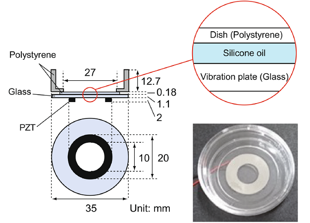
The proposed approach uses an ultrasound piezoelectric transducer glued to the bottom of a circular glass plate. A regular polystyrene cell culture dish is placed on top of the glass plate, with silicone oil used as a material coupling to efficiently transmit ultrasonic vibrations to the culture surface. This simple system is then placed inside a dedicated chamber that controls temperature, humidity, and CO2 concentration. A function generator is used to send a sinusoidal electrical signal to the transducer.
In their study, the researchers conducted a series of experiments using the above-described setup on C2C12 cells, a well-studied mice myoblast strain that can differentiate into myotubes. Specifically, the team investigated the effects of frequency, amplitude, timing, and duration of ultrasonication on the differentiation and final orientation of the cultured myoblasts. With careful analysis using 2D fast Fourier transform on phase-contrast images, they were able to quantitatively evaluate the directionality of image features related to myotube orientation.
The experiments led to some interesting conclusions, as Prof. Koyama notes: “We observed that cell orientation distribution correlated with vibrational amplitude distribution, indicating that cells grew in a direction that minimized variations in vibrational displacement along their long axes.” In other words, C2C12 matured into myotubes arranged circumferentially relative to the center of the plate. Notably, these circumferential cell orientations were observed even at radii greater than 4 mm where the vibrational displacement caused by the ultrasonic transducer was negligible. The researchers suggested that this could be due to cell-to-cell communication transiently established between myoblasts before they fuse into myotubes.
Taking their proposed approach one step further, the team conducted real-time PCR and immunostaining to shed further light on the effects induced by ultrasonication. They observed that ultrasonicated cells exhibited higher levels of differentiation-related genes, along with characteristic morphological changes associated with differentiation. Together, these results suggest that ultrasonication promotes differentiation of myoblasts into myotubes. As a result, ultrasound stimulation could serve as a promising alternative to or complement chemically induced differentiation methods.
Overall, the proposed ultrasound-based strategy offers a simple and effective method for controlling the orientation of cultured muscle cells. However, more thorough testing may be necessary to reach an optimal protocol. “The screening of different ultrasound conditions, including different frequency, output intensity, timing, and duration, will be crucial for establishing an optimized method for ultrasound-induced myotube formation in the future,” remarks Prof. Koyama.
Further refinements in the proposed strategy could pave the way to innovative solutions to current challenges in regenerative medicine using cell sheets. With continued progress, this technology could lead to life-changing clinical procedures in the coming decade, particularly in the applications of cultured skeletal muscle tissues.

Overview of the experimental setup for ultrasonication-directed myoblast orientation.
Using a simple yet effective technique, researchers from Doshisha University could harness ultrasound vibrations generated by a piezoelectric transducer to control the orientation of cultured myoblasts. After optimizing different timing, frequency, and intensity configurations, they developed a method to induce myoblasts to differentiate into circumferentially aligned myotubes.
Image courtesy: Daisuke Koyama from Doshisha University, Japan
Image license: CC BY-NC-ND 4.0
Image link: https://www.nature.com/articles/s41598-024-77277-x
Usage Restrictions: Credit must be given to the creator. Only noncommercial uses of the work are permitted. No derivatives or adaptations of the work are permitted.
Reference
| Title of original paper | Control of myotube orientation using ultrasonication |
| Journal | Scientific Reports |
| DOI | 10.1038/s41598-024-77277-x |
Funding information
This study received no funding.
EurekAlert!
https://www.eurekalert.org/news-releases/1066629
Profile

Daisuke Koyama obtained his Master’s and PhD degrees from Doshisha University in 2002 and 2005, respectively. After a few years at Tokyo Institute of Technology, he returned to his alma mater as Associate Professor in 2012 and was promoted to full Professor in 2018. Prof. Koyama specializes in ultrasonic electronics, nonlinear acoustics, and the medical applications of ultrasound and has over 100 publications to his name on these subjects. He is also a distinguished member of several academic societies, including the Acoustical Society of Japan and the Acoustic Wave Device Technology Research Consortium.
Daisuke Koyama
Professor, Faculty of Science and Engineering Department of Electrical Engineering
Media contact
Organization for Research Initiatives & Development
Doshisha University
Kyotanabe, Kyoto 610-0394, JAPAN
CONTACT US
